
Atomic and Molecular science encompasses the study of atoms, ions, and molecules, including their structure and properties, their interactions with light, and their interactions with other forms of matter. Optical science is a part of many disciplines ranging from biology to astronomy. It is also an interesting subject in its own right, encompassing a variety of phenomena associated with understanding and control of light. My research has deep roots in Atomic & Molecular Physics and Optical Science. Optics continues to play a critical role in my condensed matter and medical research programs. Selected research from my group in Optical Sciences and Atomic & Molecular Physics is described below.
Optical Sciences
Optical science pervades nearly all of the research in the Yodh lab. In some cases novel optical techniques are developed and applied to understand questions in condensed matter physics and/or biomedical optics. In other cases light interactions are of central interest as a phenomenon.
 Light Forces (Laser Tweezers)
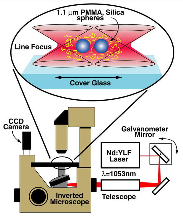
Optical tweezers exploit optical gradient forces to trap dielectric particles in three dimensions near the waist of a strongly focused laser beam. We use optical tweezers to micro-manipulate small particles in complex fluid environments. Our primary application of this tool is oriented towards measurements of colloidal interactions. For example, we recently developed a ‘line’ optical tweezer in order to trap and track the relative positions of two colloidal particles in suspension. Data are collected by video microscopy using the microscope and optical tweezer system represented schematically in the figure below. Some of the information about colloidal interactions gleaned from these experiments is described in Colloidal Interactions.
We scan the focus from side to side, rapidly enough so that the particle cannot follow the trap and so responds to the time averaged optical field. The effective optical trapping potential is thus confined to a line within the sample, hence the name line optical tweezer. In this trap, micron-size colloidal spheres are free to diffuse in one dimension, along the scan direction, while being strongly confined in the other two perpendicular directions. The particles act as if they are threaded on a frictionless rod of light. The effective interaction potential of the two spheres is related to the probability of finding the two spheres with particular center-to-center separations. A typical experiment will measure the colloidal interaction potential with and without added perturbers (e.g. perturbers could be polymers in solution, rods, nanoparticles, etc.). This procedures enables us to elucidate perturber effects with femto-newton forces and over ~10 nanometer length scales.
Two particles trapped in the line tweezer.

Many particles trapped in the line tweezer.
Confocal Microscopy
Confocal microscopy is a popular imaging methodology in biology that effectively rejects scattered light so that high fidelity near-surface (i.e. within a few hundred microns) microscope images of tissues can be obtained. It has recently found application in soft condensed matter. For example, high magnification, high-contrast three-dimensional imaging of index matched fluorescent colloidal particles (nominally 0.3 to 3.0 micron-diameter) is readily possible with current optical microscope and particle systems. The full three-dimensional imaging capability enables experimenters to track a variety of phenomena ranging from colloidal crystallization and glass formation to defect characterization and motion. In the figure we show a confocal microscopy image of 20 layers within the interior of a ~30-40 layer FCC colloidal crystal. Also shown is a reconstruction of the lattice based on the coordinates of the particles determined from the microscope images.
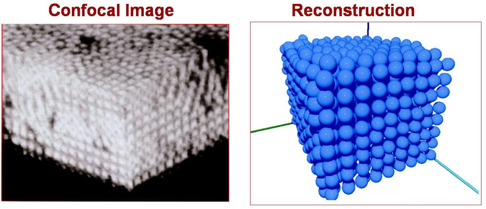
Confocal image (left) and reconstruction (right) of micron-size PMMA particles in an organic solvent. The crystals were made by templated entropic self-assembly
In vivo GRIN lens based and two-photon fluorescence microscopy
We are developing a range of portable microscopes that can be used for observation and tracking of the neuronal architecture of the superficial layers of small animal cortex. We have recently demonstrated epi-illumination GRIN lens based, three-dimensional in vivo fluorescence microscopy of neural activity using voltage sensitive dyes. We are also developing a portable 2-photon microscope for high spatial and temporal resolution studies of voltage sensitive dyes in neural networks.

Schematic of cortical activation experiment with GRIN lens.
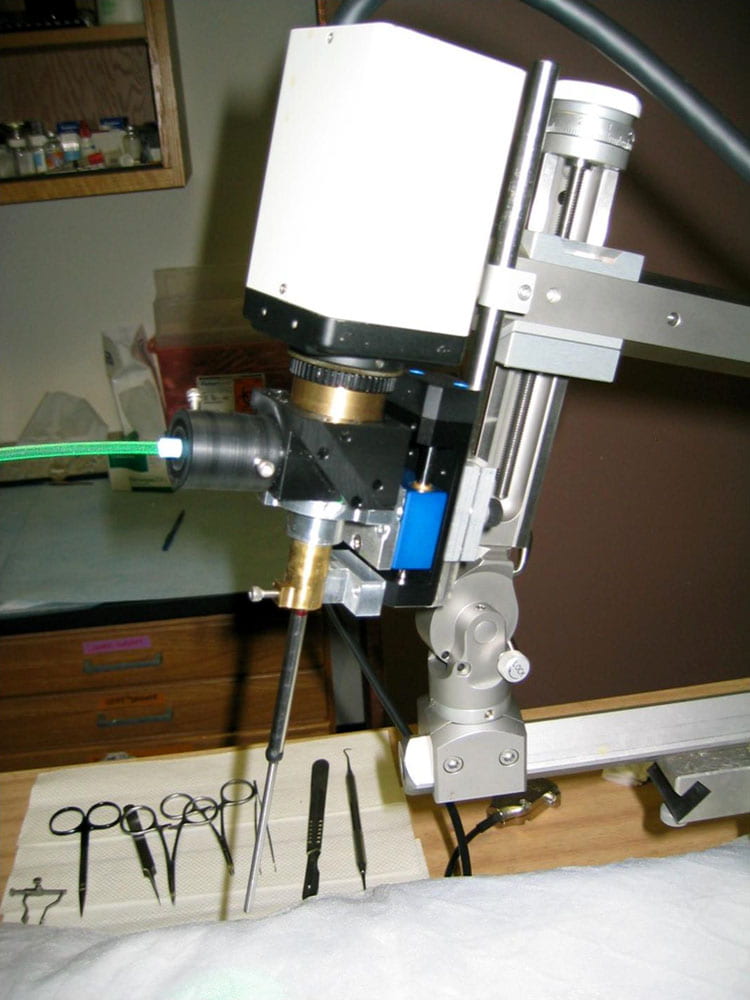
GRIN lens microscope.
 Center for Advanced Imaging and Micromanipulation
Yodh directs a MRSEC user optical microscopy facility. The Center for Advanced Imaging and Micromanipulation (CAIM) is housed within the Laboratory for Research on the Structure of Matter (LRSM) at PENN. The centerpiece of the workstation are confocal microscopes which enable three dimensional imaging and tracking of soft materials. Other capabilities include standard visualization tools such as differential interference and phase contrast, and material manipulators such as micropipettes and laser tweezers. The center also develops new capabilities for research interests of MRSEC faculty including microrheology, 2-photon microscopy and 2-photon optical lithography.
Scattering (Visible Light, X-Ray, Neutron)
The Yodh lab has an apparatus for static and dynamic (quasi-elastic) light scattering. At various times our research has required use of scattering techniques to probe complex fluids samples. For example we have used visible light and x-rays to study the properties of colloidal crystals grown by convective assembly and colloidal crystals immersed in thermotropic liquid crystals. We have also employed neutron scattering to characterize suspensions of single-wall carbon nanotubes.
Diffusing Light Optics, Imaging, and Spectroscopy
 Many materials are visually opaque because photons traveling within them are predominantly scattered rather than absorbed. Some common examples of these highly scattering media include white paint, foam, mayonnaise, and human tissue. Indeed, anyone who has held a flashlight up to his or her hand will notice some of this light is transmitted, albeit after experiencing many scattering events. Light travels through these materials in a process similar to heat diffusion. The Yodh group is deeply immersed in an interdisciplinary field of optical research whose central aim is to understand light transport in highly scattering materials, and whose second aim is to use this knowledge in order to characterize a variety of turbid media.

Physics: Our group has elucidated the physical properties of diffuse photon density waves, demonstrating refraction, diffraction, and fluorescent reemission of these disturbances. We have investigated the diffusion of light through highly scattering liquid crystals and studied its resultant dynamical fluctuations; recently we have measured anisotropy in weak localization of light from liquid crystals. In a series of studies of complex fluids we have employed diffusing-wave spectroscopy and variants to directly measure the interspecies hydrodynamic coupling between unlike particles in concentrated binary particle suspensions, and to measure Brownian dynamics on the earliest timescales. We have demonstrated that simple diffuse light transmission measurements may be used as a function of wavelength and particle concentration to provide quantitative information about colloid structure factors, and we have introduced the correlation diffusion equation, which generalized traditional diffusing-wave spectroscopy, providing a simple mathematical approach for understanding how motional fluctuations are impressed upon the spectrum of diffusing light in heterogeneous turbid media.
Biomedical Optics: The use of light to investigate the interior of human and animal tissues has grown enormously in recent years, in part as a result of advances in our fundamental understanding about light transport in highly scattering materials, and in part as a result of technological innovations in optics. We are involved in nearly all aspects of this field including the science of diffuse optical imaging, development of algorithms for data analysis, and development of understanding about the fundamental limits of diffuse optics. Current physiological applications of diffuse light imaging and spectroscopy in our lab include functional imaging and quantification of brain activation and metabolism, study of muscle function and physiology, diffuse optical tomography of breast, and characterization of tumors and tumor responses to therapy.
Nonlinear Optical Scattering, Spectroscopy and Microscopy
We are interested in understanding and using the nonlinear optical responses of materials. Three-wave mixing spectroscopy (e.g. second-harmonic generation (SHG) and sum-frequency generation (SFG)), for example, is useful for probing electronic structure and dynamics of surfaces and interfaces; we have made extensive use of this methodology in research on buried solid-solid interfaces.
More recently we have explored the nonlinear responses of colloidal particles. In particular, we carried out the first experimental measurements of Second Harmonic (SH) angular radiation patterns due to colloids. We also built a scanning SHG microscope, and have used it to study the responses of single colloidal spheres on a flat substrate.
Third-order optical nonlinearities also give rise to fascinating phenomenology and application. We have recently become interesting in two-photon absorption microscopy as a means to measure neuron dynamics in brain. To this end we have built two-photon microscope for wide-field, fast (millisecong) investigation of neuron networks in animal models, and we have carried out an preliminary studies which measure the two-photon cross-sections of various voltage-sensitive dyes.
Optical Information Storage, Laser Physics, New Mid-infrared Sources
During the 1980s Yodh was involved in initial demonstrations of optical information storage with photon echoes. The basic idea of this approach was to read and write information using the broad spectral responses of ensembles of atoms and molecules. Thus information was not only stored at a particular spatial location, but also at particular spectral locations within the ensemble. Yodh also studied how optical elements in dye lasers modified performance, and developed a new solid-state high-repetition-rate picosecond mid-infrared light source for vibrational spectroscopy. The latter scheme was based on difference-frequency generation, mixing fiber-optic compressed green light pulses, with picosecond dye laser pulses in AgGaS2 crystals.
Atomic & Molecular Physics
During the 1980s Yodh worked on atomic physics problems. This research included investigation of novel photon echo effects in gases , studies of ‘soft’ coherence preserving (i.e. velocity-changing) collisions, investigation and control of ‘dressed’ atom-photon states, exploration about how strong light fields affect collisional relaxation, and two-photon excitation of the 1s-2s transition in muonium atoms.
At PENN Yodh has carried out a variety of spectroscopy measurements. For example, the group has studied the dynamics of simple molecules (e.g. CO) on simple surfaces (e.g. Cu[111]), and has measured the optical cross-sections and anisotropy of single-wall carbon nanotubes.