A schematic of our photo-caged dimerizer is shown below, followed by two videos demonstrating organelle motility induced by uncaging in live cells.
See Zhang et al. 2017 and Chen et al. 2021 for development of new dimerizers and their application to optogenetic control of kinetochores.
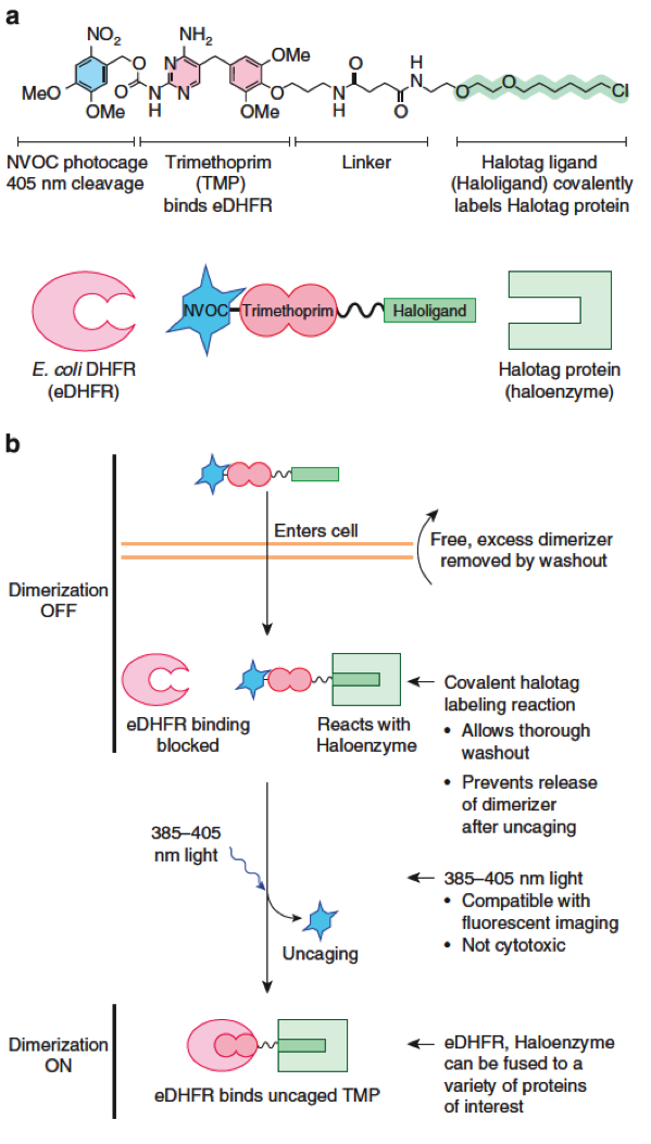
Modular design of a photocaged dimerizer. (a) Chemical structure and schematic of the dimerizer and its two receptors: eDHFR and the Halotag protein. (b) Schematic of light-induced dimerization. The modular design facilitates design of new functions, such as a different ligands or photocages or a photo-cleavable linker. From Ballister et al. 2014.
The video shows photo-activated peroxisome transport towards either microtubule plus ends (top) or minus ends (bottom). Panels show GFP-labelled peroxisomes (left) and mCherry-labeled kinesin light chain 1 (KLC1, right top) or dynein adaptor (BICD, right bottom). The dimerizer was uncaged globally by whole cell illumination. From Ballister et al. 2015.
video: localized kinesin recruitment to peroxisomes
The video shows photo-activated peroxisome transport towards microtubule plus ends, which are oriented towards the cell periphery. Panels show GFP-labelled peroxisomes (left), mCherry-labeled kinesin light chain 1 (KLC1, middle), and two color merge (right). A 405 nm laser was used to target kinesin motors to peroxisomes in the boxed region by locally uncaging the dimerizer.